What Brown Saw and You Can Too
III. Jiggly
When the seeds Douglas had shipped out arrived at the Horticultural Society in early spring 1826, they came under the purview of John Lindley (1799-1865).(1) Lindley had been mentored by Brown: in 1818, Brown gave Lindley a job that lasted a year and a half working in Banks's herbarium.(2) In 1821, the Horticultural Society leased 33 acres in Chiswick for an experimental garden.(3) The next year, Sabine hired Lindley to be assistant secretary of the garden, to superintend the collection of plants and their propagation.
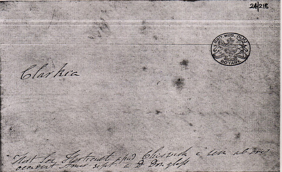 |
| First page of Brown's notes (Botany Library, Natural History Museum, London) |
The Natural History Museum in London (which spun off from the British Museum in 1881) has an extensive collection of Brown's papers. In box 24 of Brown's ``Slips Catalogue," sheet #224 is labeled Clarkia.(4) Directly underneath, Brown's (not always legible) handwriting reads Hort Soc (Horticultural Society) Horticult (illegible) Chiswick (illegible), and the next line reads occident (western) Amer (illegible) by D Douglas. Thus, Brown certifies that his Clarkia
pulchella flowers came directly from the Horticultural Society's garden. Flowers or seeds could be distributed to Fellows of the Society, but Brown was not one. Therefore, he likely received the flowers flowers privately from John Lindley.(5)
Sheet #224 contains entries dated June 12, 1827 and June 13, 1827. The first entry begins by describing the pollen:(6)
The grains of Pollen are subspherical or orbiculate-lenticular (circular-lens shaped) with three equidistant more pellucid and slightly projecting points so that they are obtusely triangular. ...
Here are two figures, one showing the the pollen viewed under a microscope, while the other is an electron
 |
| Clarkia pulchella pollen imaged by a microscope at x400 |
microscope picture of the pollen. It looks vaguely like a pinched tetrahedron with the longest dimension around 100 microns(7) and ``pores" at three vertices.
The entry turns to the contents of the pollen:
The fovilla or granules fill the whole orbicular disk but do not extend to the projecting angles. They are not spherical but oblong or nearly cylindrical. \& the particles have a manifest motion. This motion is only visible to my lens which magnifies 370 times. The motion is obscure but yet certain. ...
Thus began the research that resulted in Brown's wonderfully discursive paper,(8) dated July 30, 1828, and entitled:
A brief Account of Microscopical Observations made in the Months of June, July and August 1827, on the Particles contained in the Pollen of Plants; and on the general Existence of active Molecules in Organic and Inorganic Bodies.
It was first privately published as a pamphlet, treated as a preprint and given to various colleagues. However, it was
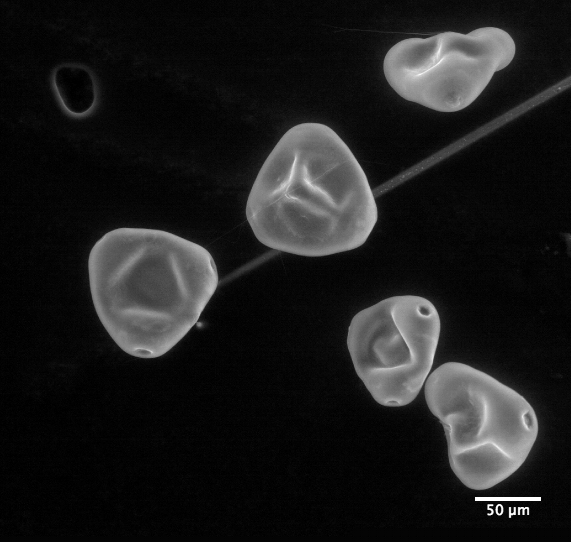 |
| Clarkia pulchella pollen imaged by an electron microscope |
published in September. It is a superb example of a researcher of unusual capability and energy delineating his thought processes. Some of its 53 paragraphs shall be treated here in some detail, especially the first nine that describe Brown's interaction with Clarkia pulchella and the start of a broader investigation.
A. Brown's Microscopes
The paper begins with a description of his microscope in the first paragraph:
The observations, of which it is my object to give a summary in the following pages, have all been made with a simple microscope, and indeed with one and the same lens, the focal length of which is about 1/32 of an inch.
Brown expands in a footnote:
This double convex Lens, which has been several years in my possession, I obtained from Mr. Bancks, optician, in the Strand. After I had made considerable progress in the inquiry, I explained the nature of my subject to Mr. Dollond, who obligingly made for me a simple pocket microscope, having very delicate adjustment, and furnished with excellent lenses, two of which are of much higher power than than that above mentioned. ...
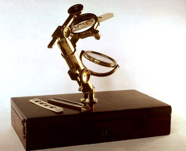 |
| A Bancks microscope owned by Brown (Linnean Society) |
However, he added that he only used the Dollond microscope ... in investigating several minute points.
A well known rule of thumb is that a near object is best seen at a distance of 10 inches. This puts the magnification Brown used at approximately 10/f=320, which is not far from Brown's own estimate of x370 cited above.
The whereabouts of this lens is not known. There does exist a pocket microscope of Brown's made by Bancks, in a box of dimensions less than 1"x2"x5". Upon Brown's death, Bennett gave this microscope ... which he was in the daily habit of using at the museum ...(9) to a mutual friend, and it has ended up at the Linnean Society. It has a complete set of lenses, the strongest of which has magnification x170. Bennett gifted another Bancks microscope, used by Brown at home, whose strongest lens has magnification x160: this is now in the museum at Kew Gardens. These are all the extant microscopes that can definitively be traced to Brown. There is also a pocket microscope at the University Museum of Utrecht, made by Dollond, with highest power lenses x330 and x480 magnification that associated documents suggest bears a relationship to Brown's Dollond microscope.
The Linnean Society microscope's x170 lens has been conjectured by Ford to be the one Brown used for his
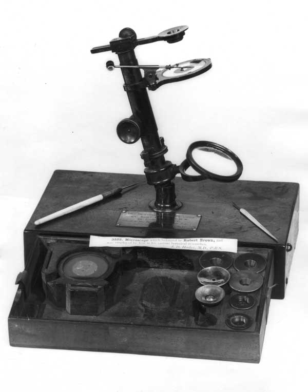 |
| Another Bancks' microscope owned by Brown (Kew Gardens) |
Brownian motion observations(9)(10), and the microscope at Utrecht is thought to be essentially identical to Brown's microscope made by Dollond. Ford proposed that Brown meant the working distance of the lens (the distance between the front of the lens and the viewed object) when he stated that the focal length was 1/32 inch. Ford also surmised that the above-mentioned extant microscopes represent Brown's full collection. If so, since the x170 lens is the strongest Bancks lens extant, it is the best candidate. In addition, Brownian motion can be observed with it,(11) albeit of milk fat globules. Indeed, as Brown asserted in his footnote, the x170 lens is much less powerful than the Utrecht x330 and x480 Dollond lenses.
However, these conjectures are doubtful.
The x170 lens (which therefore has a focal length of
1/17 inch) was measured by Ford to have a working distance of 1.5mm=1/17inch, not 1/32inch.(10) [Regardless, there must be some mistake. Half the lens thickness is the difference between the working distance and the focal length of the lens (which is essentially the object distance, for a magnifying glass). If both these numbers are 1/17 inch, this implies that half the lens thickness is 0!]
Moreover, Brown had many microscopes. Upon his death, the Gardener's Chronicle magazine reported that at least 9 microscopes of his were sold, some made by Bancks.(12)
Brown likely had two Dollond lenses of power much larger than x370, as he said in his footnote. The French botanist Alphonse de Candolle (1806-1893) visited Brown in 1828. He wrote to his famous botanist father that Brown had showed him the motion of granules from pollen, and added(13):
For that he only works with the simple lenses. But it is true that the lenses of English manufacture are as strong as many compound microscopes, because they magnify up to 800 and 1000 times. Mr. Brown has had 30 or 40 made by Dollond and other famous opticians and he chooses from them 5 or 6 in number, with which he usually works. He obtains thus the effect of an ordinary microscope with the clarity and the reliability of a simple lens.
This is supported in a remark contained in an addendum by Brown entitled Additional Remarks on Active Molecules written a year later.(8) Brown says that the new work described there
... employed the simple microscope mentioned in the Pamphlet as having been made for me by Mr. Dollond, and of which the three lenses that I have generally used, are of a 40th, 60th and 70th of an inch focus.
Thus, he says he has lenses of power x 600 and x 700, which agrees with his footnoted remark, two of which are of much higher power than the x370 lens.
Brown was the most astute microscopist of his day, and known to be extremely cautious with his statements. We believe he should be taken at his word: he used a x370 lens.
These are remarkably small lenses, with surface radii, thickness and diameter comparable in size to the focal length. Such lenses are like those of Leeuwenhoek (1632-1723)---a delightful recent paper describes grinding such a lens.(14)
Brown apparently preferred simple microscopes rather than compound microscopes. Charles Darwin (1809-1882) visited Brown in 1831, just before the voyage of the Beagle, to consult about what microscope to take. He wrote in
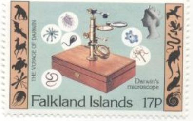 |
| Bancks microscope used by Darwin on the Beagle voyage |
his ``Life and Letters," I saw a good deal of Robert Brown ... He seemed to me to be chiefly remarkable for the minuteness of his observations and their perfect accuracy. .... He was advised by Brown to take a Bancks single lens microscope on the voyage, which he did. This microscope is at Darwin's home, Down House in Kent.
The way to construct a compound microscope that was superior to a single lens was not well known at the time, because of spherical aberration. Joseph Jackson Lister (1786-1869) (father of the surgeon Joseph Lister who instigated antiseptic operations, after whom the mouthwash Listerine was named) discovered how to minimize spherical aberration in compound microscopes, by appropriately separating lens elements. He commissioned construction of such a microscope in 1826, but only published the concept in 1830.(15)
As is discussed in detail later in this paper, a single lens, with appropriate choice of the exit pupil, can have negligible spherical aberration. In addition, a single lens microscope is more portable. Darwin only replaced his Beagle microscope, which served the dual purpose of observation and dissection, by two microscopes, a compound microscope in 1847 and a dissecting microscope of his own design in 1848. Concerning the latter, he wrote to a friend: ... I have derived such infinitely great advantage from my new simple microscope, in comparison with the one which I used on the Beagle ... . I really feel quite a personal gratitude to this form of microscope & quite a hatred to my old one.(16)
B. Observing Clarkia pulchella
The second paragraph mentions a paper Brown had published in 1826, which
... led me to attend more minutely than I had before done to the structure of the Pollen, and to inquire into its mode of action on the Pistillum ...
The pistil, the female part of a flower, consists of a vase-like object called the style, containing at its bottom the ovules
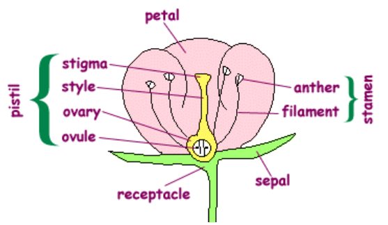 |
| Flower parts |
(immature seeds containing eggs) and a structure on top called the stigma. Others conjectured that, when a pollen grain sticks to the stigma, the grain releases the particles it contains, and these somehow travel down through the style to fertilize the ovules. In his third paragraph, Brown expresses doubts respecting the mode of action of the pollen in the process of impregnation.
As explained in the fourth and fifth paragraphs, he had the idea to look into this too late in the year, past the time of flowering:
It was not until late in the autumn of 1826 that I could attend to this subject; and the season was too far advanced to enable me to pursue the investigation. Finding, however, in one of the few plants then examined, the figure of the particles contained in the grain of pollen clearly discernible, and that figure not spherical but oblong, I expected with some confidence to meet with plants in other respects more favorable to the inquiry, in which these particles, from peculiarity of form, might be traced through their whole course ... .
I commenced my study in June 1827, and the first plant examined proved in some respects remarkably well adapted to the object in view.
Thus Brown explains his selection: among a number of flowers apparently chosen by chance, Clarkia pulchella pollen clearly contained oblong particles.
For what follows, note that the male part of a flower, the stamen, consists of two parts. There is the anther, which is a sack in which pollen grains develop; it sits on a stalk called the filament, which conveys nutrients from the flower to the anther. When the pollen is ripe, it is released because the anther bursts, splitting longitudinally (in most cases), a process called dehiscence.
The sixth paragraphs launches the investigation.
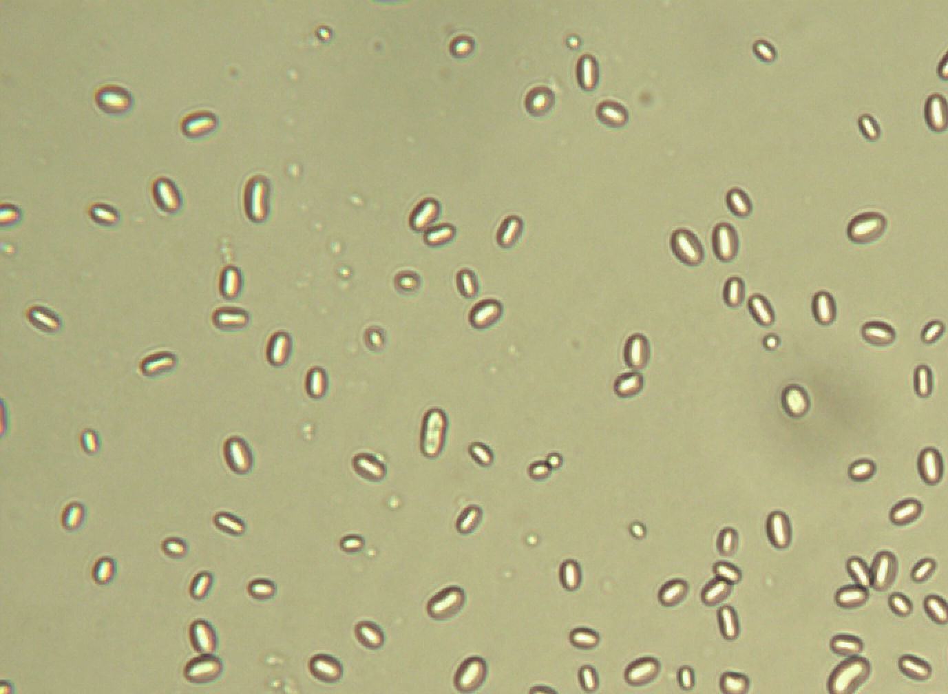 |
| Amyloplasts and (a few) spherosomes from Clarkia pulchella |
This plant was Clarckia pulchella, of which the grains of pollen, taken from antherae fully grown before bursting, were filled with particles or granules of unusually large size, varying from 1/4000th to about 1/3000th of an inch in length, and of a figure between cylindrical and oblong, ... . While examining the form of these particles immersed in water, I observed many of them very evidently in motion ... . In a few instances the particle was seen to turn on its longer axis. These motions were such as to satisfy me, after frequently repeated observation, that they arose neither from currents in the fluid, nor from its gradual evaporation, but belonged to the particle itself.
This is the first kind of particle Brown observes, whose length he estimates at about 6 to 8 microns. This is worth noting, since observations with modern equipment discussed later in this paper give these particles shorter lengths. The difference shall be attributed to the alteration of the image by his lens, as mentioned earlier.
In the seventh and eighth paragraphs, he notes the existence of a second kind of particle;
Grains of pollen of the same plant taken from antherae immediately after bursting, contained similar subcylindrical particles, in reduced numbers however, and mixed with other particles, at least as numerous, of much smaller size, apparently spherical, and in rapid oscillatory motion.
These smaller particles, or Molecules as I shall term them, when first seen, I considered to be some of the cylindrical particles swimming vertically in the fluid. But frequent and careful examination lessened my confidence in this supposition; and on continuing to observe them until the water had entirely evaporated, both the cylindrical particles and spherical molecules were found on the stage of my microscope.
C. Seeing Brownian Motion
We emphasize here that Brown was not observing the pollen move. He was observing much smaller objects, which reside within the pollen, move. This is well known---see for example the excellent pedagogical article by Layton.(17) Nonetheless, statements that Brown saw the pollen move are rife.(18)
A Clarkia pollen is approximately 100microns across.(7) As we shall shortly show, that is too large for its Brownian motion to be readily seen. However, fortunately for Brown, the contents of the pollen are just the right size for their motion to be conveniently observed.
D. Observing Pollen Of Other Plants
In paragraph 9, Brown starts to look at the pollen of other plants, to see if their contents are similar and behave similarly. First, he looks at plants which have a similar classification. In the family Oenothera (evening primrose), Clarkia is a genus and C. pulchella is a species. Another genus in the same family is Onagraceawhich Brown calls Onagrariae:
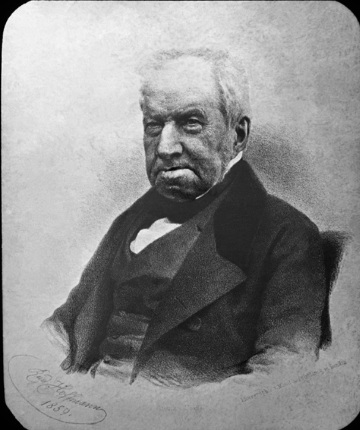 | |
| Robert Brown (photo published after his death) |
In paragraph 10, Brown remarks that this
... reduction in that of the cylindrical particles, before the grain of pollen could possibly have come in contact with the stigma, --- were perplexing circumstances in this stage of the inquiry, and certainly not favorable to the supposition of the cylindrical particles acting directly upon the ovulum; an opinion which I was inclined to adopt when I first saw them in motion. ...
In paragraph 11 he is off and running, looking at a variety of flowering plants: In all these plants particles were found, which in the different families or genera varied in form from oblong to spherical, having manifest motions similar to those already described ... In a great proportion of these plants I also remarked the reduction of the larger particles, and a corresponding increase of the molecules after the bursting of the antherae ...
Prior to discussing the next paragraph, we should emphasize that, so far, Brown had not observed the particles or granules moving while they were within the Clarkia pulchella pollen grain. As he says in paragraphs 6 and 8, he observed them moving in water. Unfortunately, he doesn't say how they manage to get out of the pollen grain after the grains are put in water. As will be discussed in more detail iin a later section, pollen grains in water---in vitro---may burst open, the contents streaming out under pressure (called turgor). (What happens naturally---in vivo---will be discussed there too.) Moreover, the particles within Clarkia pulchella pollen seem to be too packed together to move. And, we have observed that the fluid in which they are packed is so viscous that their motion is impeded when they do emerge. However, paragraph 12 says:
In many plants, belonging to several different families, but especially to Gramineae, the membrane of the grain of pollen is so transparent that the motion of the larger particles within the entire grain was distinctly visible; ... and in some cases even in the body of the grain in Onagrariae.
So, Brown was able to see particles move within some pollen, but he does not specifically include Clarkia pulchella. Sometimes Brown is said to have observed particles moving within the pollen, and the implication is that this was what Brown first observed, which is incorrect.(19)
The next two paragraphs consider plants with varied kinds of pollen but similar results. Then comes paragraph 15:
Having found motion in the particles of all the living plants which I had examined, I was led next to inquire whether this property continued after the death of the plant, and for what length of time it was retained.
Paragraph 16 reports that, from plants dried or preserved in alcohol, for a few days, to a year, to more than twenty years, to more than a century, the pollen ... still exhibited the molecules or smaller spherical particles in considerable numbers, and in evident motion, ... .
He next has the idea to look at plants that reproduce by spores: mosses and the horsetail (Equisetum). He finds within the moss spores, and sitting on the surface of the Equisetum spores, ... minute spherical particles, apparently of the same size with the molecule described in Onagrariae, and having equally vivid motion on immersion in water ; ... .
E. Observing Organics
Then, as described in paragraph 19, an accident occurred. On bruising a spore of Equisetum, ... which at first happened accidentally, I so greatly increased the number of moving particles that the source of the added quantity could not be doubted. This leads him to bruise ... all other parts of those plants ... , with the same motion observed.
Therefore, the motion had nothing to do with plant reproduction. He says:
... My supposed test of the male organ was therefore necessarily abandoned.
From this comes a hypothesis. The naturalist George-Louis Leclerc, Comte de Buffon (1707-1788), had proposed an atomic-style hypothesis, that there are elementary "organic molecules" (hence Brown's name for the smaller particles he observed) out of which all life is constructed. Brown signs onto this in paragraph 20:
... I now therefore expected to find these molecules in all organic bodies: and accordingly in examining the various animal and vegetable tissues, whether living or dead, they were always found to exist; and merely by bruising these substances in water, I never failed to disengage the molecules in sufficient numbers to ascertain their apparent identity in size, form, and motion, with the smaller particles of the grains of pollen.
Paragraph 21 contains this charming observation:
... I remark here also, partly as a caution to those who may hereafter engage in the same inquiry, that the dust or soot deposited on all bodies in such quantity, especially in London, is entirely composed of these molecules.
He now looks at things that were once organic, gum-resins, pit coal, then fossil wood. He then thinks of mineralized vegetable remains and looks at silicified wood, with similar results. Paragraph 22 concludes:
... But hence I inferred that these molecules were not limited to organic bodies, nor even to their products.
F. Observing Inorganics
So, (paragraphs 23-32) ... to ascertain to what extent the molecules existed in mineral bodies became the next object of inquiry. .... Starting with ... a minute fragment of window-glass, from which when merely bruised on the stage of the microscope ..., he tries all kinds of minerals, rocks, and metals, even ... a fragment of the Sphinx!
... in a word, in every mineral I could reduce to a powder sufficiently fine to be temporarily suspended in water, I found these molecules more or less copiously: ...
When he looks at objects that are not spherical, such as fibers, he conjectures that they are composed of a number of molecules. He heats or burns wood, paper, cloth fiber, hair, quenches them in water and finds ``molecules" in motion.
G. Brown's Summary of Observations on Molecules
Paragraphs 33-37 summarize, with commendable caution:
There are three points of great importance which I was anxious to ascertain respecting these molecules, namely, their form, whether they are of uniform size, and their absolute magnitude. I am not, however, entirely satisfied with what I have been able to determine on any of these points.
As to form, I have stated the molecule to be spherical, and this I have done with some confidence; ...
He explains that he judged the size of bodies ... by placing them on a micrometer (a glass slide with lines ruled on it) divided to five thousandths of an inch ... .
The results so obtained can only be regarded as approximations, on which perhaps, for obvious reason, much reliance will not be placed. ... I am upon the whole disposed to believe the simple molecule to be of uniform size, ... its diameter appeared to vary from 1/15,000 to 1/20,000 of an inch.
So, with his microscope, he estimates the molecule size at from 1.7 to 1.3 microns. A footnote adds While this sheet was passing through the press .... he asked the lens maker Dollond to look at Equisetum spores, whose surface he had earlier noted released ``molecules,"
... with his compound achromatic microscope, having at its focus a glass divided into 10,000ths of an inch, upon which the object was placed; and although the greater number of particles or molecules seen were about 1/20, 000 of an inch, yet the smallest did not exceed 1/30,000th of an inch.
So, with Dollond's microscope, these particular molecules appeared smaller, mostly 1.3 microns, with some estimated at .85 microns.
Brown prudently concludes,
I shall not at present enter into additional details, nor shall I hazard any conjectures whatever respecting these molecules ...
H. Brown's Concluding Remarks
In the final paragraphs of the paper, Brown returns
... to the subject with which my investigations commenced, and which was indeed the only object I originally had in view ... , namely whether the larger particles acted upon the ovule. My endeavors, however, to trace them, ... was not attended with success ... .
He returned to this problem, with more success, a few years later (see the next section). The paper ends with establishing his priority. He notes: The observations, of which I have now given a brief account, were made in the months of June, July and August, 1827. He mentions people to whom he showed the phenomenon (he soon traveled to Europe, and demonstrated it there) and people who had made related observations in the past (the phenomenon was first seen by Leeuwenhoek, and remarked upon by many later microscopists, but fell short of his results in some way .
Brown issued an addendum the following year,(8) Additional Remarks on Active Molecules,
... to explain and modify a few of its statements, to advert to some of the remarks already made, either on the correctness or the originality of the observations, and to the causes that have been considered sufficient for the explanation of the phenomena.
He rejects the notion that the molecules are animated, he regrets having introduced hypotheses such as larger objects being made out of molecules, distances himself from the notion that the molecules are identically sized, rejects some explanations of the motion. He says they are
... motions for which I am unable to account.
He describes an experiment designed to put to rest the idea that it is evaporating water, or interaction among the particles, which produces the motion, He shakes a mixture of oil and water that has previously been filled with particles, obtaining small drops of water surrounded by oil, some of which contain only one particle, and notes that the motion is unabated and continues indefinitely since the water does not evaporate.
He concludes once again by ... noticing the degree in which I consider those observations to have been anticipated, and discussing other people's earlier work.
-------------------------------------------------------------------------------------------------
1) A biography of John Lindley is available at the orchid information web site http://www.orchids.co.in/: select orchidolologists.
2) D. J. Mabberley, Jupiter Botanicus: Robert Brown of the British Museum (J. Cramer, Braunschweig 1985). The biographical details given in this paper are almost exclusively from this authoritative source.
3) The Horticultural Society garden in Chiswick was part of the 655 acre estate of the lessee, William Spencer Cavendish (1790-1858), the 6th Duke of Devonshire, with a gate to the garden installed for the Duke's use. The physicist Henry Cavendish (1731-1810) was related, a grandson of the 2nd Duke. What remains of this estate, Chiswick House on 68 acres, a fine example of Palladian architecture, is being restored and can be visited (http://www.chgt.org.uk). More of the acreage exists as a Chiswick public park, Dukes Meadows (http://www.dukesmeadowstrust.org/). However, the Horticultural Society garden is no more, replaced by city streets.
4) We are indebted to David Mabberley for ``strongly" suggesting that the slips catalog be looked into, and to Armando Mendez, of the Botany Library of the Natural History Museum, for finding this and mailing us a copy.
5) We are indebted to Brent Elliot, Historian of the Royal Horticultural Society, for the following information. The society does not have any surviving plant receipt books for the garden at Chiswick until the 1840s, so one cannot track Clarkia pulchella into or out of the garden. Lindley was in overall supervision of the gardens, with Donald Munro as head gardener, and a staff of under-gardeners. It was common for botanists to share plants of interest.
6) The complete entries for both dates are reproduced in J. Ramsbottom, Journ. of Bot. 70, 13, (1932).
7) E. Small, I. J. Bassett, C. W. Crompton, and H. Lewis, \textit{Pollen Phylogeny in Clarkia}, Taxon 20, 739 (1971) contains detailed dimensional measurements on many species of Clarkia, including pulchella.
8) R. Brown, Edinburgh New Philos. Journ. 5 358, (1828) and Philos. Mag. 4 161, (1828). This, and the addendum published a year later, Edinburgh Journ. Sci 1 314, (1829), can be downloaded from the New York Botanical Gardens, at http://sciweb.nybg.org/science2/pdfs/dws/Brownian.pdf.
9) B. J. Ford, Single Lens: the Story of the Simple Microscope (Harper and Row, New York 1985), pp. 152-153.
10) B. J. Ford, Microscopy 34, 406, (1982); The Linnean 1,12, (1985). These describe the loving restoration of the Linnean Society microscope, with the first paper giving quantitative measurements on the lenses. See also B. J. Ford, Infocus 15, 18 (2009).
11) B. J. Ford, http://www.brianjford.com/wbbrowna.htm. This web site also contains a photograph of the Bancks-made Linnean Society microscope of Brown's, as well as a comparison of a view with a single lens and a compound microscope of the time, and a short video of Brownian motion of milk fat droplets. http://www.microscopy-uk.org.uk/dww/home/hombrown.htm also has a video of milk fat droplets undergoing Brownian motion, of sizes .5 to 3$\mu m$, with tips on how to duplicate the observation.
12) Mabberley, Ibid, p. 389.
13) This is a translation from the French. The original appears in Mabberley, Ibid, pp. 270-271.
14) H. Loncke, ``Making a von Leeuwenhoek microscope lens," Microscope 138 (April, 2007): this is an online article available at http://www.microscopy-uk.org.uk/mag/indexmag.html?http://www.microscopy-uk.org.uk/mag/artapr07/hl-scope.html.
15) J. J. Lister, Phil. Trans. Roy. Soc. London, 120, 187, (1830).
16) W. A. Newman in History of Carcinology (CRC Press, 1993), F. Truesdale, ed., p. 357, available at Googlebooks.
17) D. Layton, Journ. Chem. Ed. 42, 367 (1965).
18) A casual on--line search turns up many examples. J. Bernstein Am. Jour. Phy. 74, 863 (2006), in a fine, authoritative assessment of Einstein's 1905 Brownian motion paper and its impact, nonetheless writes on p.85, about Brown: ``He noticed that if pollen grains of a few ten-thousands of a centimeter were suspended in water, they executed an incessant jigging motion." The size is right, but it is not the Clarkia pollen size; Einstein year website, http://www.einsteinyear.org/facts: ``In 1827 the biologist Robert Brown noticed that if you looked at pollen grains in water through a microscope, the pollen jiggles about."; Announcement of Gauss Prize (2006) for Kiyoshi Ito, http://www.mathunion.org/Prizes/Gauss/index.html. ``In Ito's case, this way begins with a look into a microscope showing pollen grains or dust particles in water moving around in an erratic way."; T. Spencer, power point lecture ``Random Walk from Einstein to the Present" at the educational arm of the Institute for Advanced Study at Princeton, http://pcmi.ias.edu/, type in Brownian motion: ``Around 1827 Brown made a systematic study of the “swarming motion� of microscopic particles of pollen."; Wikipedia Dictionary, http://en.wiktionary.org/wiki/Brownian, ``1. Pertaining to botanist Robert Brown (1773-1858), who investigated the movement of pollen suspended in water."; http://www.wisegeek.com/what-is-brownian-motion.htm, ``As a botanist, Brown first observed the effect in pollen floating in water, where it is visible with the naked eye." ; Univ. of St. Andrews, School of Mathematics and Statistics, http://www-groups.dcs.st-and.ac.uk/~history/Biographies/Ito.html, ``Brown, a botanist, discovered the motion of pollen particles in water."; http://physicsworld.com/cws/article/print/21146, ``Most of us probably remember hearing about Brownian motion in high school, when we are taught that pollen grains jiggle around randomly in water due the impacts of millions of invisible molecules."; http://www.math.utah.edu/~carlson/teaching/java/prob/brownianmotion/1/, ``In 1827 the English botanist Robert Brown noticed that pollen grains suspended in water jiggled about under the lens of the microscope, following a zigzag path like the one pictured below."
19) B. J.Ford, The Microscope 40, 235 (1992) verified that Brown could have seen motion with his Linnean x170 lens, writing: ``The Clarkia pollen was obtained from anthers of C. pulchella at the Botanical Garden at Cambridge University, and pollen specimens from other species within the Oenotheraceae were also utilized. Exactly as Brown recorded, the experiments were carried out in the month of June and the pollen grains were mounted in water after removal from pre-dehiscent anthers. ... The phenomenon of Brownian movement was well resolved by the original microscope lens. Within the pollen grains, ceaseless movement could be observed." However, while the author saw the contents of the pollen, he did not see them undergoing Brownian motion, as this appears to suggest. Rather, with this lens, he observed the Brownian motion of similarly sized milk fat droplets (private communication from B. J. Ford). Presumably, what was meant is that ``ceaseless movement could have been observed" with that lens. Perhaps this led to the erroneous statement in an article reporting on this work, Science News 142 (Aug. 15), 109 (1992): ``Each grain contains a thick liquid ... When Brown looked inside the pollen grains with his microscope, he could see tiny particles, each about 1 micron across, suspended in the liquid and constantly in motion."